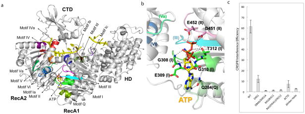
Figure 4. SF2 Helicase dissected.
(a) Mapping of the common SF2 motifs and Cas3-specific motifs onto the ATP-bound Cas3 structure. (b) Close-up of the ATP-binding interactions. Motifs are shown in parenthesis. More details are in Fig. S8. (c) Helicase region mutagenesis done using the same procedure as in Fig. 2c.

