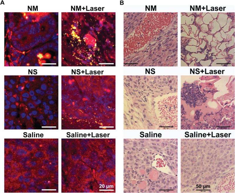
Figure 5.
Histopathology of tumor sections extracted from mice intravenously injected with gold nanoparticles or control saline solution. (Left panel) Fluorescence staining combined with dark field microscopy: cell nucleus is stained with DAPI in blue; cell cytoplasm stained with Alexa Fluor® 594 in red and gold nanoparticle can be observed as yellow bright spots due to the nanoparticle scattering in the dark field mode. (Right panel) Hematoxyline and Eosine (H&E) staining. The cell morphology in all non-treated tumors, specially the nucleus, is observed intact. However, after laser treatment in presence of gold nanomatryoshkas or nanoshells the cell morphology is disrupted.

