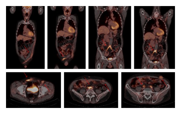
Figure 1.

Coronal and axial view of fused 18F-FDG PET/CT images. In this particular case, a 67-year-old male patient underwent an aorto-bi-iliac bypass 16 years ago. After 3 years the bypass was revised because of an occlusion. Five years later the second bypass also occluded and an axillobifemoral bypass was constructed. Unfortunately this bypass also occluded twice. Patient is admitted to the hospital because of pain and redness at the level of the axillobifemoral bypass probably due to infection. 18F-FDG PET/CT scanning showed increased FDG uptake at the level of both occluded bypasses, the aorto-bi-iliac bypass and the axillobifemoral bypass. Arrows are pointing at increased FDG uptake in both bypasses. Ultimately, no bacteria were cultured.
