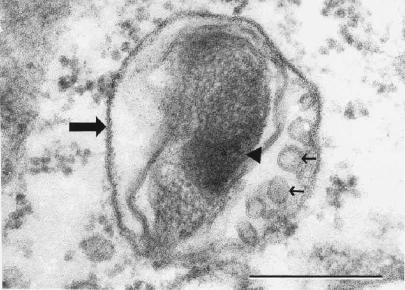
FIG. 7.
Electron photomicrograph demonstrating a damaged intravacuolar rickettsia with the morphological characteristics of early destruction (large arrow, vacuolar membrane). Note the large rickettsial nucleoplasmic electron density (arrowhead) and rickettsial cell wall membrane blebs (small arrows). Anti-OmpB antibody-treated rickettsiae after 24 h in an SVEC endothelial cell are shown. Bar, 0.25 μm.

