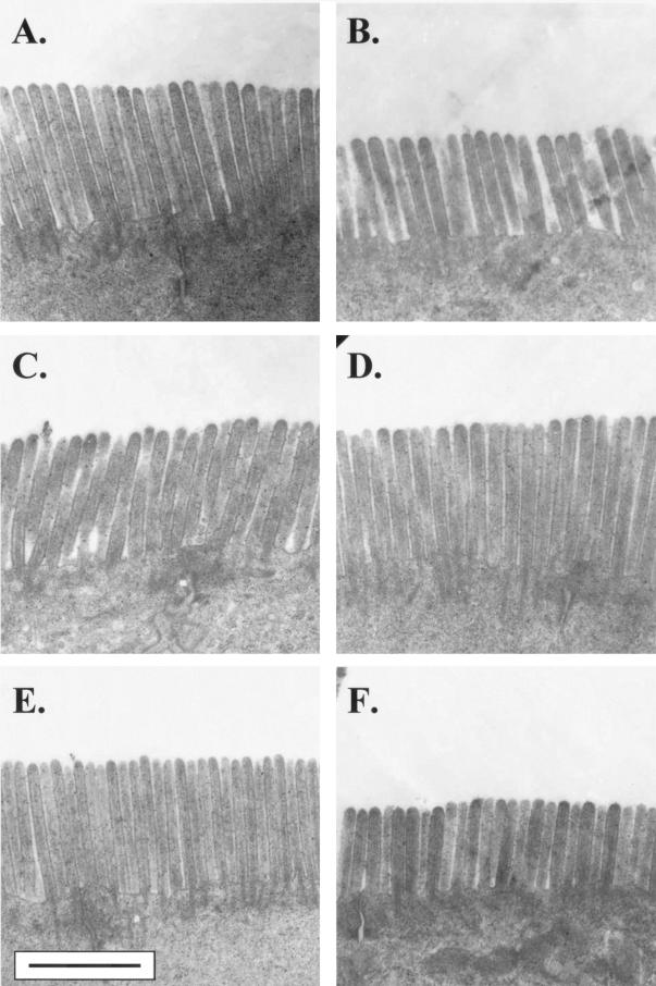
FIG. 1.
Representative transmission electron micrographs of the jejunal microvillous brush border from naive mice which received lymphocytes from donors infected with Giardia (B, D, and F) or from sham-inoculated CDs (A, C, and E). In separate experiments, mice received either whole SMLN lymphocytes (A and B), enriched SMLN CD4+ T cells (C and D), or enriched SMLN CD8+ T cells (E and F). Brush border shortening, when present, was diffuse and was seen at sites of trophozoite colonization as well as in other areas. Bar equals 1 μm for all micrographs.

