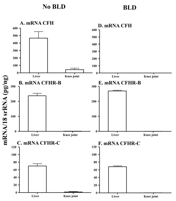
FIGURE 3.
mRNA expression levels of CFH, CFHR-B and CFHR-C analysis from liver and knee joint of CfhTg/mCfh−/− mice without and with BLD. Only liver and knee joint from 12-month-old CfhTg/mCfh−/− chimeric mice (with BLD) and age-matched littermate B6 mice (with no BLD) were analyzed. All RNA samples were analyzed in duplicate by qRT-PCR. A. CFH mRNA was present in liver and knee joints of B6 mice with no BLD using primers from exon #3. B. CFHR-B was present in liver and knee joints of B6 mice with no BLD. C. CFHR-C was present in liver and knee joints of B6 mice with no BLD. D. No mouse fH mRNA was detected in liver and knee joint of CfhTg/mCfh−/− chimeric mice with BLD using primers described previously (Ufret-Vincenty et al., 2010) but mRNA for CFH was present using primers from exon #3 (data not shown). E. CFHR-B mRNA in liver and knee joints of CfhTg/mCfh−/− mice with BLD. F. CFHR-C mRNA in liver and knee joint of CfhTg/mCfh−/− mice with BLD mice. All data presented in these graphs, as Mean ± SEM, were from CfhTg/mCfh−/− mice with BLD (n = 5) and B6 mice (n = 5) with no BLD. These experiments were repeated three times. *p-value < 0.05 compared to liver

