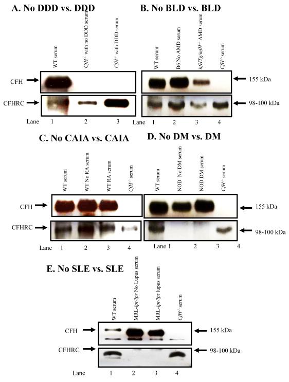
FIGURE 7.
Western blot analyses showing the presence of CFHR-C in mouse serum with and without DDD, BLD and CAIA and also showing the universal absence of mice with and without DM and SLE. Sera from WT C57BL/6 and fH−/− mice were used as a positive and a negative control for CFH and CFHR-C, respectively. Mouse serum was analyzed by using 8% Trisglycine SDS-PAGE under reducing conditions. Immuno-blotting was done using a primary anti-CFH and a secondary HRP-conjugated antibody. A. Serum was analyzed from WT (fH+/+) and fH−/− mice with no DDD and with DDD. Top panel (lanes 1, 2 & 3): Serum from WT (fH+/+) mice, 6-week-old fH−/− mice with no DDD and 40-week-old fH−/− mice with DDD. No CFH is present in fH−/− mice with DDD, as expected. Bottom panel (lanes 1, 2 & 3): Serum from WT (fH+/+) mice with no DDD and fH−/− mice with DDD. A band of ~98-100 kDa of CFHR-C was present in serum from WT, and in fH−/− with no DDD and with DDD, respectively. B. Sera from B6 mice with no BLD and from CfhTg/mCfh−/− chimeric mice with BLD were analyzed. Top panel (lanes 1, 2 & 3): A band of CFH was present in serum from WT, with no BLD and with AMD mouse. Bottom panel (Lanes 1, 2, 3 & 4): CFHR-C was present in serum from WT, B6, CfhTg/mCfh−/− and fH−/− mouse. C. Sera from WT C57BL/6 mice with no CAIA, at day 0, and WT C57BL/6 mice with CAIA, at day 10, were analyzed. Top panel (lanes 1, 2 & 3): A band of CFH was present in the sera from WT, with no CAIA and with CAIA mice. Lane 4. Absence of CFH in serum from fH−/− mice, as expected. Bottom panel (Lanes 1, 4): A band of CFHR-C was present in sera from WT, with no arthritis, with arthritis and fH−/− mice. D. Sera from NOD mice with no DM, and NOD mice with DM were analyzed. Top panel (lanes 1, 2 & 3): A band of fH ~155 kDa was present in serum from WT, no DM and DM mice. Lane 4. Absence of CFH in serum from fH−/− mouse as a negative control. Bottom panel (Lanes 1, 4): A band of ~98-100 kDa of CFHR-C was present in serum from WT and fH−/− mice. Lanes 2, 3. CFHR-C was absent in serum from mice with no DM or with DM. E. Sera from MRL-lpr/lpr without disease and MRL-lpr/lpr with disease mice were analyzed. Top panel (lanes 1, 2 & 3): A band of CFH was present in sera from WT, MRL-lpr/lpr (12 Wks) and MRL-lpr/lpr (23 Wks) mouse. Lane 4. Absence of CFH in the sera from fH−/− mice as expected. Bottom panel (lanes 1): A band of CFHR-C was present in sera from WT mouse. Occasionally, twin bands were also seen at ~96-100 kDa and these may be variants of CFHR-C. Lanes 2 & 3. No CFHR-C protein was present in sera from MRL-lpr/lpr mice with and without lupus, respectively. Lane 4. CFHR-C protein was present in sera from fH−/− mice even in the absence of CFH. Western blots using each serum were repeated at least three times and results were highly reproducible.

