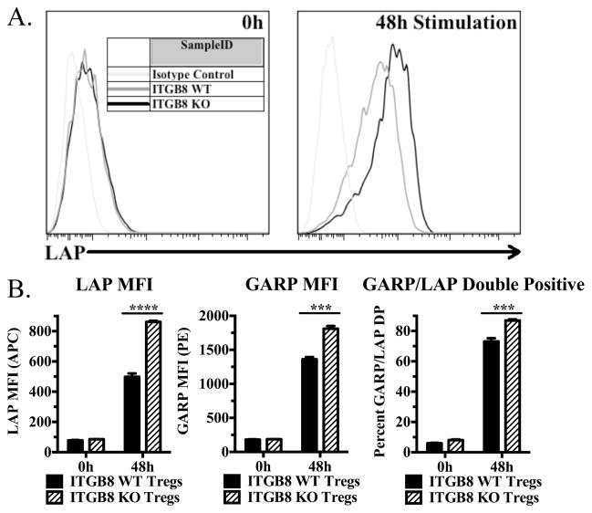
FIGURE 3.
Activated Itgb8-deficient Tregs accumulate latent TGF-β1 on their surface. (A and B) Freshly isolated cells from pooled lymph nodes and enriched CD4+ cells from Itgb8 conditional knockout mice (CD4-CRE) or CRE- littermates were stimulated for 48 h with plate-bound anti-CD3 and IL-2, then stained for CD4, Foxp3, LAP (or IC), and GARP (or IC). (A) Representative histograms for LAP (latent TGF-β1) from CD4+Foxp3+ cells. (B) Graphical representations indicating LAP and GARP MFI on CD4+Foxp3+ cells as well as percentage of LAP and GARP double positive cells.

