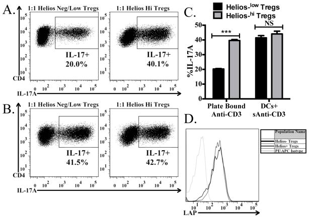
FIGURE 6.
Helios low/- cells are less efficient at producing biologically active TGF-β1. Naive CD45.1+ OT-II cells from Rag1 −/− mice were activated by plate-bound (A) or soluble ani-CD3+ splenic DCs in the presence of IL-6 and preactivated Helios+ or Helios-/low cells from Helios (GFP)/Foxp3 (RFP) double reporter mice. After 4 days, cells were reactivated with Cell Stimulation Cocktail and Protein Transport Inhibitors and then stained for CD4, CD45.1, CD45.2, and IL-17A. The percentage of IL-17A+ cells derived from the CD4+CD45.1+CD45.2− cells in the culture are shown in each panel. (C) Bar graph indicates the average (± SD) of duplicates within the experiment from (A) and (B). (D) Tregs were activated for 48 hours, then stained for CD4, Foxp3, Helios, and LAP. Histograms shown represent the expression of LAP on CD4+Foxp3+Helioshi cells and CD4+Foxp3+Helios−/low cells, as compared to the isotype control.

