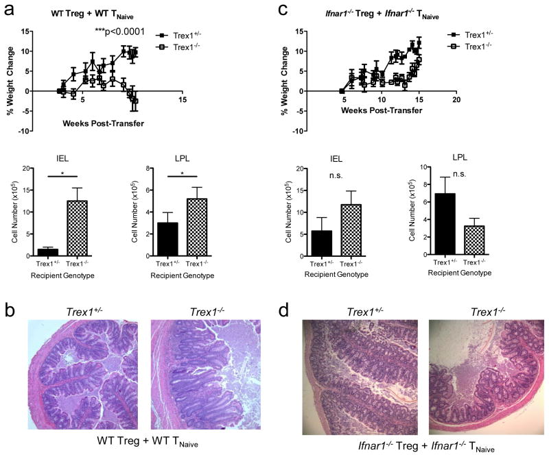
Figure 1. IFNαR signaling in T cells is required for immune dysfunction in Trex1−/− mice.
a) Top: percent weight change in Rag2−/−Trex1+/−(“Trex1+/−“, black squares) and Rag2−/−Trex1−/−(“Trex1−/−“, open squares) mice at various time points after co-transfer of WT CD45.2+CD4+CD25+ Treg and WT CD45.1+CD4+Foxp3GFP−CD45RBhi TNaive cells. Bottom: absolute number of intraepithelial (IEL) and lamina propria lymphocytes (LPL) in the colons of Rag2−/−Trex1+/− (black) and Rag2−/−Trex1−/−(checkered) recipient mice at time of sacrifice. b) Representative H&E staining of cross-sections of intermediate to distal colon from Rag2−/−Trex1+/− and Rag2−/−Trex1−/− recipients of WT Treg and WT TNaive cells at time of sacrifice. c) Top: percent weight change in Rag2−/−Trex1+/−(“Trex1+/−“, black squares) and Rag2−/−Trex1−/−(“Trex1−/−“, open squares) mice at various time points after co-transfer of Ifnar1−/− CD45.2+CD4+CD25+ Treg and Ifnar1−/− CD45.1+CD4+Foxp3GFP−CD45RBhi TNaive cells. Bottom: absolute number of intraepithelial (IEL) and lamina propria lymphocytes (LPL) in the colons of Rag2−/−Trex1+/− (black) and Rag2−/−Trex1−/−(checkered) recipient mice at time of sacrifice. d) Representative H&E staining of cross-sections of intermediate to distal colon from Rag2−/−Trex1+/− and Rag2−/−Trex1−/− recipients of Ifnar1−/− Treg and Ifnar1−/− TNaive cells at time of sacrifice. Statistical significance was determined using unpaired two-tailed Student’s t-test. Data are representative of two independent experiments with 3–4 mice per group. *, p<0.05; **, p<0.005; ***, p<0.0001.

