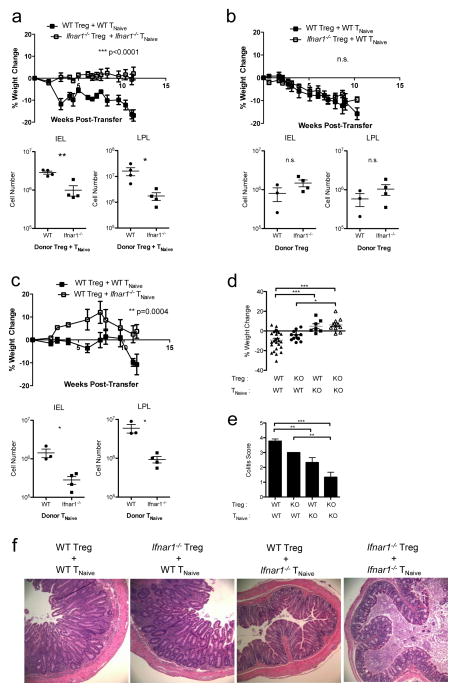
Figure 4. IFNαR signaling in effector, but not regulatory, T cells is required for colitis development in Trex1−/− mice.
a–c) Top: percent weight change in Rag2−/−Trex1− /− mice at various time points after co-transfer of WT Treg + WT TNaive cells (black squares) or co-transfer of: (a) Ifnar1−/− Treg + Ifnar1−/− TNaive cells (open squares); (b) Ifnar1−/− Treg + WT TNaive cells (open squares); or (c) WT Treg + Ifnar1−/− TNaive cells (open squares). Bottom: absolute number of intraepithelial (IEL) and lamina propria lymphocytes (LPL) in the colons of the indicated Rag2−/−Trex1−/− mice at time of sacrifice. Data are representative of 2–3 independent experiments with 3–4 mice per group. d) summary of the final percent weight change at time of sacrifice in Rag2−/−Trex1−/− mice receiving the indicated WT or Ifnar1−/− (knockout, “KO”) Treg and TNaive cells. e) summary of colitis scores based on histological analysis of colon cross-sections from Rag2−/−Trex1−/− mice receiving the indicated WT or Ifnar1−/− (knockout, “KO”) Treg and TNaive cells. f) Representative H&E staining of cross-sections of intermediate to distal colon from Rag2−/−Trex1−/− recipients of the indicated Treg and TNaive cells. (d, e) Data are summarized from 8 independent experiments with 3–4 mice per group. Statistical significance was determined using unpaired two-tailed Student’s t-test (a–c) or one-way ANOVA with Tukey post-test (d, e). *, p<0.05; **, p<0.005; ***, p<0.0001.

