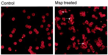
FIG. 2.
Fluorescence photomicrograph of control (left panel) and Msp-treated (right panel) neutrophils after permeabilization and incubation with rhodamine actin. The cells were treated with Msp for 1 min. Intense rhodamine fluorescence developed in the subcortical area of Msp-treated cells (arrows).

