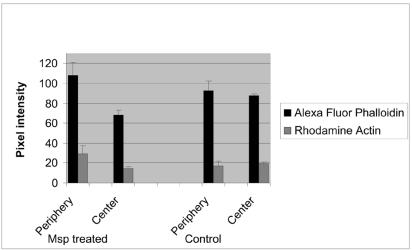
FIG. 5.
Distribution of Alexa fluor 488 and rhodamine fluorescence intensity between central and peripheral (subcortical) areas of Msp-treated and vehicle-treated control Rat-2 fibroblasts. Msp treatment increased the mean (± standard error) subcortical actin filament and actin barbed end fluorescence intensity (n = 3 experiments).

