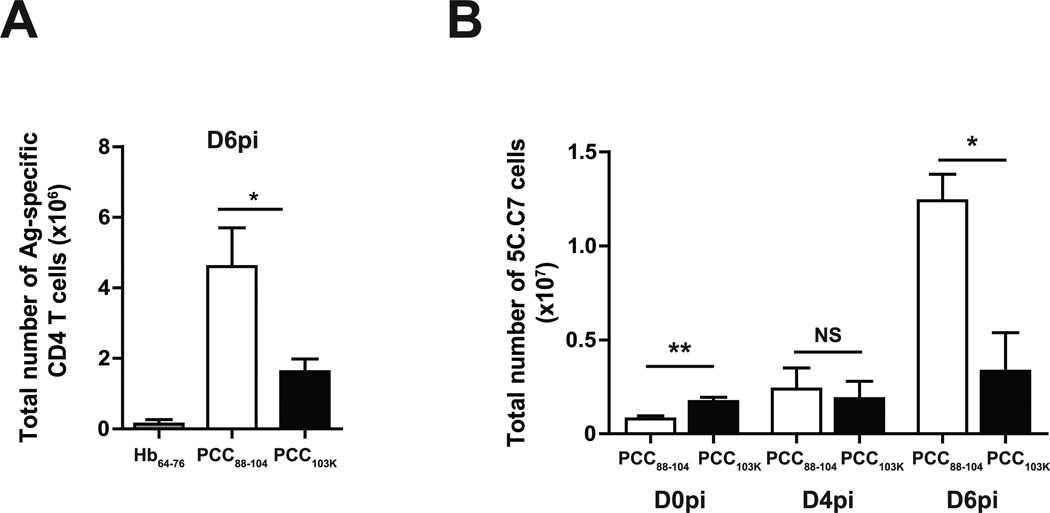
Figure 4. Better accumulation of effector CD4 T cells with low stability peptides.
(A) B10.BR mice were s.c. immunized with Hb64–76, PCC88–104, or PCC103K peptides, challenged 7 days later with WSN-MCC88–104 influenza virus, and analyzed on day 6 after challenge. Total number of Ag-specific CD4 T cells (Dapi−B220−CD8a−CD11b−Vα11+Vβ3+CD44hi) in lung. (B) Splenocytes from Thy1.2+ 5C.C7αβ TCR transgenic mice were adoptively transferred into congenic Thy1.1+ mice. Recipient mice were s.c. immunized with PCC88–104 or PCC103K peptides and challenged 7 days later with WSN-MCC88–104 influenza virus. Total number of 5C.C7 cells (Dapi−B220−CD8a−CD11b−Thy1.2+CD44hi) in lungs on days 0, 4, and 6 postinfection. Means ± SEM for at least three animals are shown. *p ≤ 0.05, **p ≤ 0.01 (unpaired Student t test). Data shown are derived from at least three independent experiments (n ≥ 3 mice per group).

