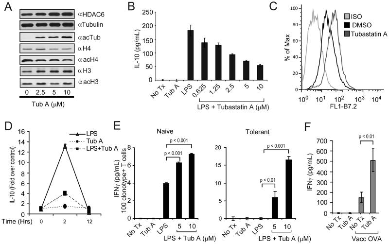
Figure 3. Phenotype and function of APCs treated with the isotype-selective HDAC6 inhibitor, Tubastatin A.
(A) PEM were treated with increasing concentrations of Tubastatin A (Tub-A) for 24 hours. Protein extracts were prepared and subjected to SDS-PAGE and immunoblotting with α–HDAC6, α-tubulin, α-acetylated tubulin, α-histone 4 (H4), α-acetylated H4, α-histone 3 (H3), α-acetylated H3 specific antibodies. (B) PEM (1×10⁵/well) were treated with LPS (1 μg/ml) alone or LPS plus increasing concentrations of Tub-A for 24 hours. Then, supernatants were collected and the production of IL-10 was determined by ELISA. (C) Expression of B7.2 on macrophages treated with Tub-A (5 μM) was determined by flow cytometry. (D) PEM (2×10⁶/well) were treated with 1ug/ml of LPS (triangles), 5μM of Tub-A (circles), or the combination thereof (squares). Cells were harvested at 0, 2 and 12 hours and total RNA was isolated. The IL-10 expression by quantitative RT-PCR is expressed as fold over non treated control cells and normalized by GAPDH expression. Data (A-D) is from a representative experiment of three experiments with similar results. (E) PEM (1×10⁵/well) were treated as in (B). Then, cells were washed and 5×10⁴ purified naïve (left) or tolerized (right) anti-HA CD4+T-cells were added to the cultures in the presence of 12.5 μg/ml of cognate HA-peptide110-120. After 48 hours, supernatants were collected and assayed for IFN-γ production by ELISA. Data is from a representative experiment of three independent experiments with similar results. (F) C57BL/6 mice were adoptively transferred with 2.5×10⁶ anti-OVA TCR transgenic CD4+ T-cells given intravenously (day zero). Animals were then treated with either Tub-A (25mg/kg) or vehicle control given intraperitoneally (ip) for 10 days (day zero to +9). On day +9 half the mice in each group were immunized s.c. with 1×10⁷ pfu of vaccOVA. Animals were sacrificed six days later and T-cells were purified from their spleens as indicated in Methods. T-cells were then cultured with splenocytes from C57BL/6 mice in the presence of 3 μg/ml of OVA323-339 peptide. After 48 hours, supernatants were collected and IFN-γ production was determined by ELISA. Data represent mean ± SE of triplicate cultures from 8 mice in each group and is representative of two independent experiments with similar results. Values for T-cells cultured without OVA-peptide were below the limit of detection.

