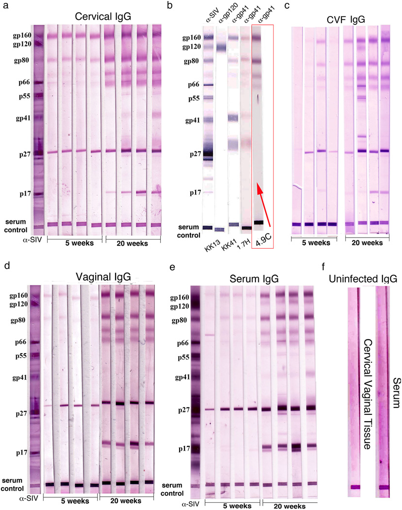
FIGURE 2.
Increased SIV-gp41 IgG antibodies correlate with the temporal maturation of protection. Each lane in the WBs represents an individual animal. (a, c, d) Increases between 5 and 20 weeks in IgG-antibodies in cervical vaginal tissue extracts or fluids reacting with oligomeric Env gp41antigens-gp160, gp80, and two other glycoproteins of lower molecular wt. Lanes marked α–SIV indicate positive control polyclonal antibody; serum control indicates that sample was loaded. (b) Rhesus monoclonal antibodies to gp41 and gp120 identify prominent Env antigen bands as oligomeric gp41. The band at ~31 kDa is thought to be a truncated species of gp41. The WB lane with a rhesus monoclonal antibody, 4.9C, which reacts identically to cervical tissue antibodies to oligomeric gp41, is indicated by a red box and arrow. (e) Similar increases and SIV-reactivities in IgG in serum. (f) Cervical vaginal tissue and serum controls, SIV-uninfected tissues.

