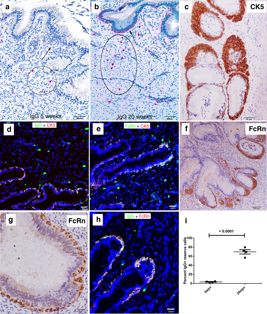
FIGURE 3.
Increases between 5 and 20 weeks in IgG+ plasma cells, and IgG+FcRn+ cervical reserve epithelium. (a, b) Red-stained IgG+ cells with plasma cell morphology (encircled) in the submucosa at 5 and 20 weeks. Arrows point to cells with epithelial morphology aligned beneath the columnar epithelium identified in (c, d) as cytokeratin 5 (CK5)+ IgG+ cervical reserve epithelium. (e) The CK5+ cervical reserve epithelium is FcRn+. (f, g) Brown-stained FcRn+ cells with identical morphology and location beneath the columnar epithelium as CK5+ cervical reserve epithelium shown in (c). (h) IgG is concentrated in the FcRn+ reserve epithelium. (i) Quantification of the relative numbers at 5 and 20 weeks of IgG+ plasma cells in the submucosa and IgG+ cervical reserve epithelium. Four animals analyzed at 5 weeks and four animals at 20 weeks.

