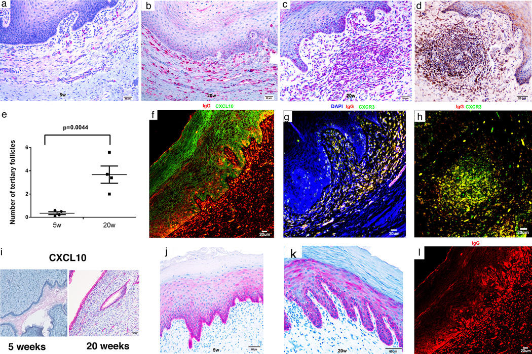
FIGURE 6.
Recruitment of plasma cells to vagina and cervix; induction of ectopic lymphoid follicles; and localization of IgG in FcRn+ basal epithelium in the lower FRT. (a,b) Increased red-stained plasma cells in the submucosa at 20 weeks compared to 5 weeks. (c–e) Increases between 5 and 20 weeks in ectopic tertiary follicles in vagina. Follicles contain red-stained IgG+ plasma cells in (c) and brown-stained gp41t-antibody+ cells in (d). (f–h). Epithelial expression of CXCL10 and recruitment of CXCR3+ plasma cells to the submucosa and ectopic follicles. In (f) CXCL10 expressing vaginal epithelium is stained green and the IgG in plasma cells is stained red. The orange appearance in the merged confocal micrograph is the result of IgG concentrated by the FcRn in CXCL10+ basal epithelium (see also j, k and l). (g, h) CXCR3+ IgG+ plasma cells in the submucosa and in ectopic follicles are stained yellow. (i) Increased expression of CXCL10 between 5 and 20 weeks in red-stained endocervical epithelium. (j,k) Mainly basal epithelial expression of FcRn at 5 and 20 weeks. (l) IgG concentrated in the FcRn+ epithelium above IgG+ plasma cells. Image corresponds to (f) but only red-staining IgG shown.

