
Figure 2.
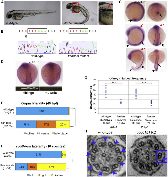
Zebrafish ccdc151 Is Expressed in Ciliated Tissues and Required for Ciliary Motility-Dependent Processes
(A) Wild-type (left) and flanders (ts272a) mutant (center and right) zebrafish embryos at 48 hr postfertilization (hpf). Right panel is a magnification of the boxed region in the center panel. Red arrow indicates pronephric cysts evident in flanders mutants.
(B) DNA sequencing chromatograms demonstrating the ccdc151 c.631T>A (p.Lys211∗) nonsense mutation in flanders mutants.
(C) In situ hybridization demonstrating wild-type ccdc151 expression in Kupffer’s vesicle at tailbud stage (black arrows, top panels) and intermediate mesoderm (black arrows), otic vesicles (red arrows), and neural tube (black arrowheads) during early (10 somites; middle panels) and late (20 somites; bottom panels) somitogenesis.
(D) In situ hybridization demonstrating loss of ccdc151 expression in flanders mutants. Progeny of flanders/+ incrosses were sorted based on ccdc151 expression and genotyped using the DraI restriction site destroyed by the c.631T>A (p.Lys211∗) mutation.
(E) Quantification of asymmetric organ positioning in embryos at 48 hpf. flanders mutants and siblings were sorted based on body curvature morphology prior to in situ hybridization. ∗∗∗chi-square p value < 10−55.
(F) Quantification of asymmetric expression of zebrafish nodal homolog Southpaw during somitogenesis. flanders mutants and siblings were distinguished based on DraI restriction site presence or absence following in situ hybridization. ∗∗∗chi-square p value < 10−9.
(G) Quantification of pronephric cilia beat frequency in flanders mutants and siblings at 48 hpf and 72 hpf. ∗∗∗Student’s t test p value < 10−16.
(H) Transmission electron microscopy of cross-sections through pronephric cilia reveals the lack of outer dynein arms in embryos injected with the ccdc151 morpholino (KD = knockdown; blue box and arrow), compared to a wild-type uninjected embryo where outer dynein arms (blue arrows) are visible. Scale bars represent 50 nm.