
Figure 3.
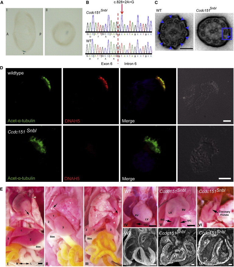
Ccdc151 Localizes to Embryonic Node Monocilia and Ccdc151Snbl Mutants Exhibit a Spectrum of Laterality Phenotypes Including Complex Congenital Heart Defects Associated with Heterotaxy
(A) Whole-mount in situ hybridization analysis of Ccdc151 in wild-type E7.5 mouse embryos shows that Ccdc151 is specifically expressed in the ventral node, marked with an asterisk in panel I showing a view from the left. Panel II shows a ventral view. Abbreviations are as follows: A, anterior; P, posterior.
(B) Sequence of the Ccdc151Snbl homozygous mutant mouse (RefSeq NM_029939) compared to a wild-type littermate shows a c.828+2A>G substitution affecting the exon 6 splice donor site, with the exon-intron boundary shown by a dashed red line and intronic sequence distinguished from exonic sequence by lower case.
(C) Transmission electron microscopy of tracheal cilia from wild-type and a Ccdc151Snbl homozygous mutant mouse reveals the lack of outer dynein arms in the mutant (blue arrowheads). Scale bars represent 0.1 μm.
(D) Air-dried tracheal airway epithelia from wild-type and Ccdc151Snbl mutant mice costained for acetylated α-tubulin (green) and the ODA component DNAH5 (red) were visualized by immunofluorescence microscopy. Nuclei were stained with DAPI (blue). In control mice, DNAH5 localized to the axonemes stained with acetylated α-tubulin (top panels), but in Ccdc151Snbl mutant airway epithelia, DNAH5 is undetectable in the ciliary axonemes (bottom panels) Scale bars represent 10 μm.
(E) Homozygous Ccdc151Snbl mutants exhibit a spectrum of laterality defects including situs solitus (I), situs inversus totalis (II), or heterotaxy (III). In I–III, heart situs is denoted by arrows, lung lobation is numbered 1–5 (white numbers), and liver lobation is numbered 1–3 (black numbers); Stm indicates stomach. Dextrocardia, inverted lung lobation, inverted liver lobation, and dextrogastria are seen in the situs inversus totalis mutant (II) as compared to the normal visceral organ situs observed in the situs solitus mutant (I). A mutant with heterotaxy (III) exhibits levocardia with normal heart orientation and lung lobation but inverted liver lobation and dextrogastria. Analysis of the cardiovascular anatomy of two Ccdc151Snbl mutants with heterotaxy revealed one with congenital heart defects (V) comprising dextrocardia with duplicated inferior vena cava (IVC) and a ventricular septal defect (VSD; VIII) and another mutant heart with a coronary artery fistula spanning from the left coronary artery to the left ventricle (VI, IX). For comparison, the heart from a normal control animal with situs solitus is shown (IV, VII). The R-L double arrow in (I) indicates right-left orientation, which is the same for all panels. Abbreviations: LV, left ventricle; RV, right ventricle.