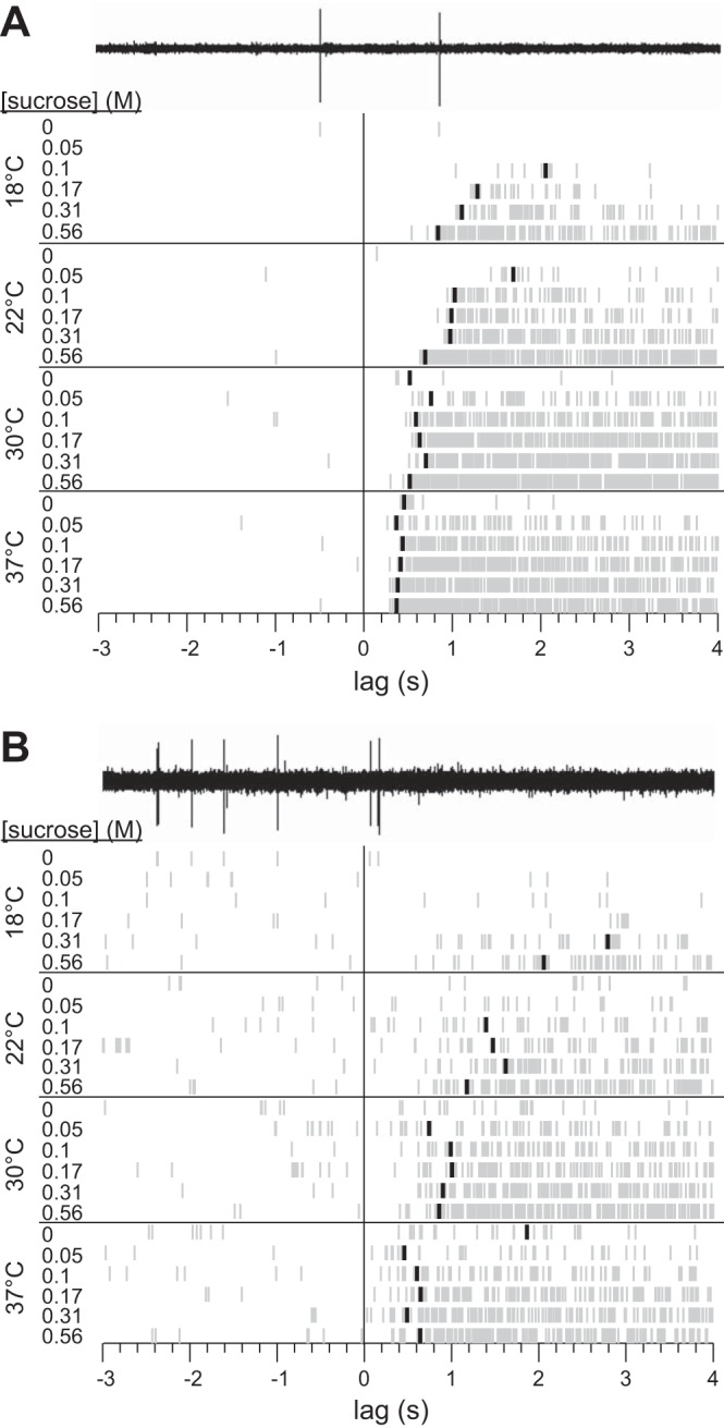
Fig. 4.

Rastergrams for 2 separately recorded S-type neurons (A and B) showing detection of latency to first spike across 24 unique temperature-concentration combination trials for sucrose. The electrophysiological sweep recorded for 0 M sucrose at 18°C is shown for each cell to demonstrate conversion of neurophysiological data to raster spikes. A blackened raster spike on a trial represents the time during stimulus delivery when the firing rate of the neuron became unusually high compared with the average prestimulus firing rate of the cell (see materials and methods). The absence of a blackened spike indicates that no significant elevation in firing was detected for that trial.
