Fig. 1.
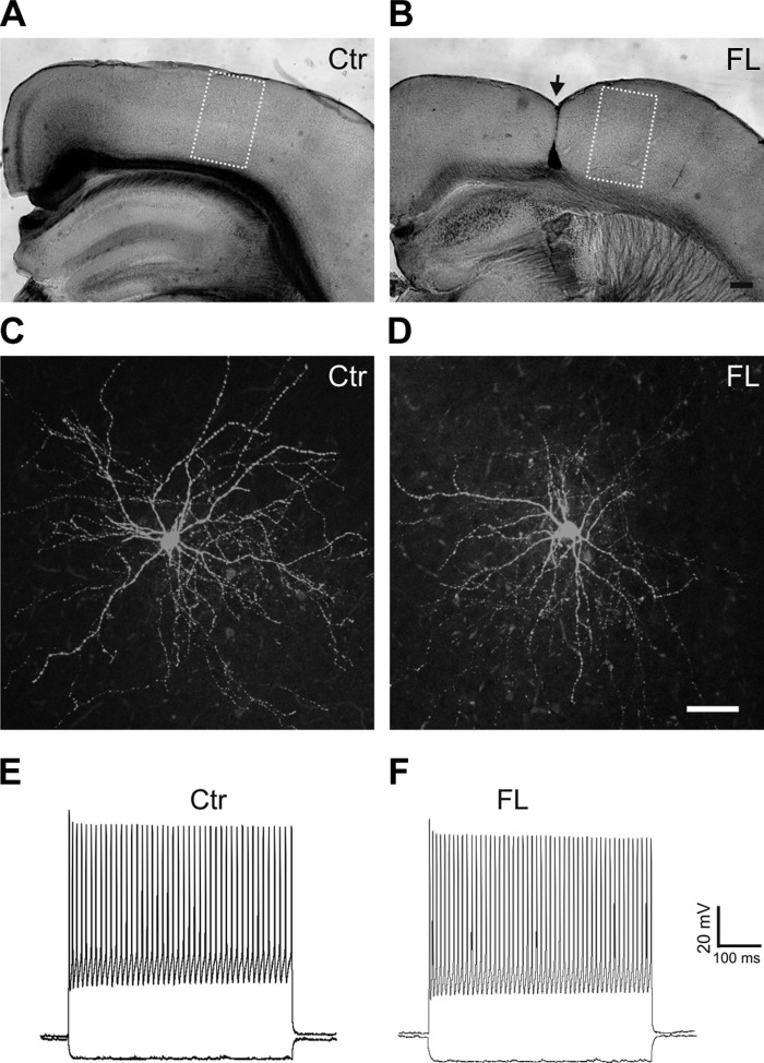
Green fluorescent protein (GFP)-expressing fast spiking (FS) interneurons in the control (Ctr) and freeze lesion (FL) cortex. A and B: images of fixed coronal cortical sections from control (A) and FL (B) mice. The yellow rectangles delineate uncaging areas that cover ∼500–600 × 1,000–1,200 μm of the cortex. The location of the FL lesion is marked with arrow in B. C and D: confocal images of biocytin-filled GFP-expressing neurons in layer V of control (C) and FL (D) neocortical slices processed with avidin D fluorescein reveal typical morphology of FS interneurons with smooth multipolar dendrites and dense local axonal arbors in cortical layer V. E and F: depolarizing current pulses in GFP neurons evoke high-frequency trains of fast action potentials without obvious adaptation in the control (E) and FL cortex (F). Scale bars = 200 μm in B for A and B; 50 μm in D for C and D.

