
Figure 3.
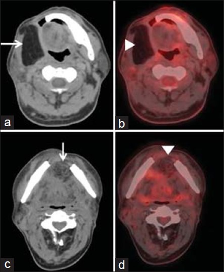
Tissue changes after surgery. Axial fused PET/CT images show myocutaneous flap with fat density used to reconstruct the defect after hemimandibulectomy (arrow in a) and mandibular symphysectomy (arrow in b) performed for buccal cancer absence of normal physiological FDG uptake in the reconstructed flap (arrowheads in a and b)