Figure 4.

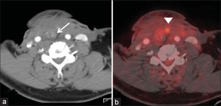
Tissue changes after surgery. Axial CT shows a reconstructed neopharynx after a laryngopharyngectomy seen as a tubular structure (arrow in a). Diffuse low grade physiological uptake is seen around the neopharynx (arrow head in b)

Tissue changes after surgery. Axial CT shows a reconstructed neopharynx after a laryngopharyngectomy seen as a tubular structure (arrow in a). Diffuse low grade physiological uptake is seen around the neopharynx (arrow head in b)