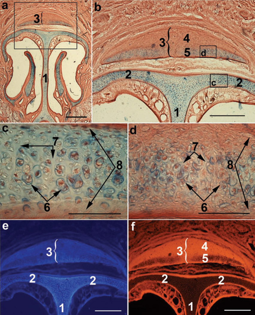
Figure 4.
Light microscopy and fluorescence of nasal cartilages of an adult rat. (a) Coronal slice of the entire nasal cartilaginous skeleton stained with Alcian blue and counterstained with Thiazine red. (b) Boxed area in (a). Boxed areas in (b) represent roof cartilage (c) and the ventral area/compartment of the dorsal nasal cartilage (d). (e), (f) Blue autofluorescence and Thiazine red fluorescence, respectively, of the area shown in (b). (1) Septum; (2) roof cartilage; (3) dorsal nasal cartilage; (4) dorsal and (5) ventral compartments of the dorsal nasal cartilage; (6) chondrocytes clustered in groups; (7) matrix; (8) perichondrium. Scale bars are 1 mm (a), 0.5 mm (b, e and f), and 0.1 mm (c, d).

