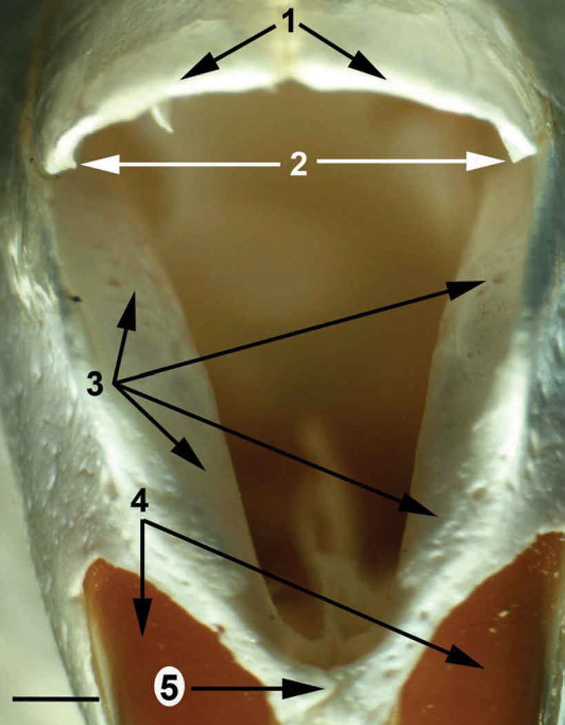
Figure 5.
Light microscopy of horizontal and coronal slices of the rat snout stained for cytochrome oxidase activity. (a) A horizontal slice cut through the snout of a 17-day-old rat at the ventral margin of the nostril. (b) Light microscopy of the coronal slices cut at the positions shown at the bottom of the panel (a). (1) atrioturbinate; (2) rostral edge of the atrioturbinate; (3) narial pads; (4) ostium of the nasolacrimal duct; (5) lateral ventral process; (6) septal Fenestrae; (9) fragments of the intraturbinate muscles; (10) the bone of the maxilloturbinate; (11) rostral edge of the premaxilla; (12) caudal part of the nasal cartilaginous skeleton. Scale bars = 1 mm.

