. 2014 Sep 9;3:e02443. doi: 10.7554/eLife.02443

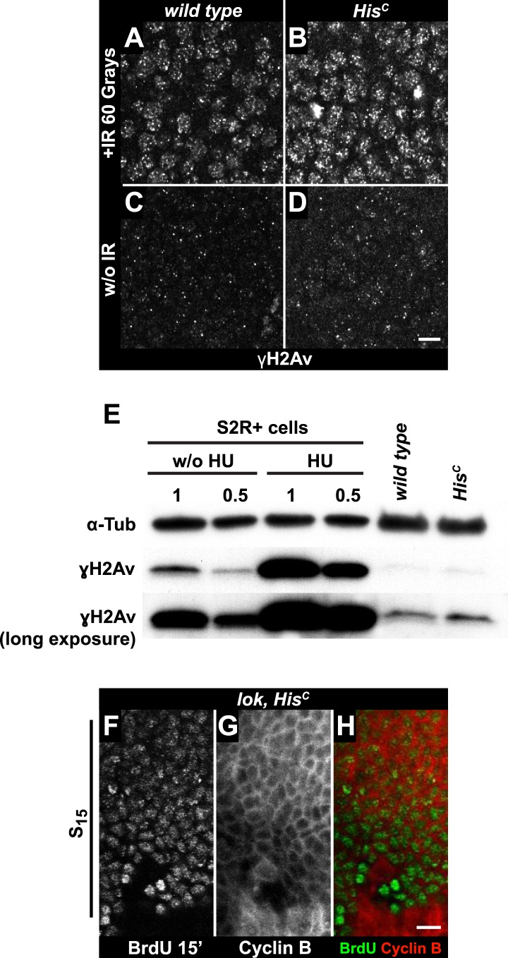
Figure 4. The cell cycle arrest of HisC mutant cells does not depend on the ATM/Chk2 DNA damage checkpoint.

(A and B) Wild type and HisC mutant cells responded to ionizing irradiation (IR) by phosphorylation of the variant histone H2Av, detected by a phosphospecific antibody against H2Av (γH2Av). (C and D) Without irradiation, HisC mutant cells did not show elevated γH2Av staining compared to wild type, indicating that mutant cells did not accumulate DNA damage. (E) Western blot for γH2Av using untreated (w/o HU) and HU-treated (HU) S2R+ cells as controls (two dilutions, 1 and 0.5) as well as HisC mutant embryos and wild type embryos at 5.5–6.5 hr AEL. α-Tubulin (α-Tub) was used as a loading control. HU treatment results in a significant increase of γH2Av, which was not observed in HisC mutant embryos. (FH) BrdU pulse labelling for 15 min and staining with antibodies against BrdU and Cyclin B. lok, HisC double mutant embryos showed a similar phenotype as HisC mutant embryos (see Figure 1F). Dorsal up in (FH), scale bars: 10 µm.

DOI: http://dx.doi.org/10.7554/eLife.02443.010

Figure 4.

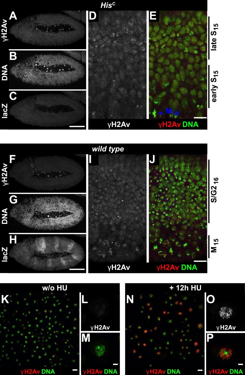
Figure 4—figure supplement 1. HisC mutant embryos show a moderate increase of γH2Av during S15 progression.

Figure 4—figure supplement 1.

HisC mutant (AE) and wild type sibling (F–J) embryos stained with antibodies against phosphorylated H2Av (γH2Av), against β-Galactosidase (lacZ) and with DAPI to detect DNA. (AC) HisC mutant embryos were identified by the lack of lacZ staining. (D and E) Late S15 cells in the dorsal epidermis of HisC mutant embryos show a moderate increase of γH2Av as compared to ventral cells that are in early S15 or still in M14 (blue arrowhead). (FH) Wild type sibling embryos of comparable age were identified based on lacZ staining. (I and J) In these embryos dorsal epidermal cells were already in S/G216. The increase in γH2Av staining was less pronounced than in HisC mutant embryos. (KP) Untreated (w/o HU; KM) and HU-treated (NP) S2R+ cells were stained for γH2Av and DNA showing an increase of γH2Av signal in HU-treated cells. Dorsal up in (AJ), anterior to the left in (AC, FH), scale bars: 100 µm (AC, FH), 10 µm (D, E, I, J), 20 µm (K and N) and 5 µm (L, M, O, P).

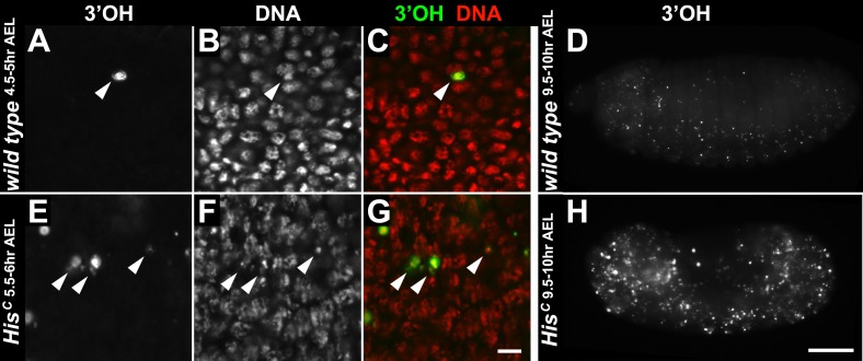
Figure 4—figure supplement 2. HisC mutant embryos do not show accumulation of DNA damage during early embryogenesis.

Figure 4—figure supplement 2.

(AH) TUNEL assays with whole mount embryos to detect free 3′OH groups of DNA, which indicate DNA damage. (AC) Wild type embryos at 4.5–5 hr AEL contained a few apoptotic cells (arrowheads). In most nuclei, free 3′OH groups were not detected. (D) Whole mount wild type embryos at 9.5–10 hr AEL showed a few apoptotic cells. (EG) HisC mutant embryos at 6.5–7 hr AEL also contained a few apoptotic cells (arrowheads). However, the level of TUNEL staining was not uniformly elevated in the mutant cells, indicating that there is no increase of DNA damage compared to wild type. (H) HisC mutant embryos die around 9.5–10 hr AEL and show an increased number of apoptotic cells. Scale bars: 10 µm (AC), (EG), 100 µm (D and H).