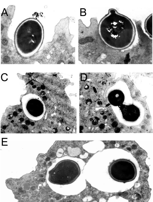
FIG. 2.
TEM of H. capsulatum cells interacting with A. castellanii. (A and B) Two separate phagocytic events at 2 h postincubation with amoebae. (C and D) Yeast cells in a membrane-bound vacuole surrounding the fungal cell 2 h after infection of the amoeba suspension with fungal cells. (E) Two individual H. capsulatum fungal cells in separate phagocytic compartments indicating two independent phagocytic events. Magnification, ×15,000 (A, B, and E) and ×12,000 (C and D).

