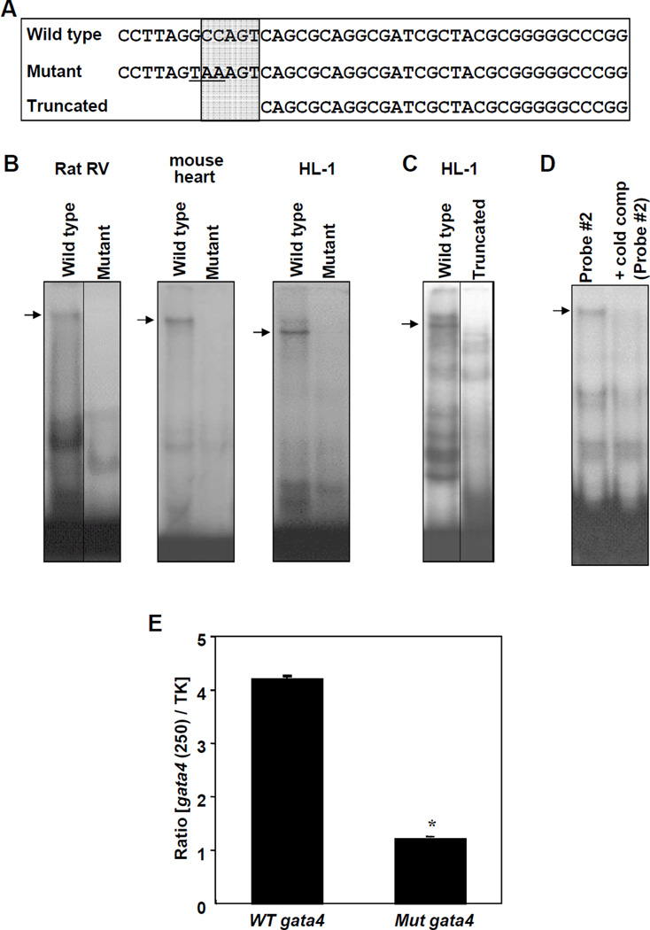
Fig. 3. Role of the CCAAT Box.
(A) Sequences of various double stranded EMSA probes. CCAAT box regions are indicated in the shaded area. (B) Nuclear extracts from rat RV (7-day hypoxia treated), mouse heart or HL-1 cardiac muscle cells were subjected to EMSA using wild type or mutant Probe #2. (C) Nuclear extracts were subjected to EMSA with wild type or truncated Probe #2. (D) Heart nuclear extracts were subjected to EMSA using 32P-labeled Probe #2 in the presence of excess cold competitor (unlabelled Probe #2). (E) HL-1 cells were transfected with the luciferase construct controlled by the 250 bp proximal region of the wild type (WT) or mutant (Mut) Gata4 promoter. Values represent means ± SEM of the ratio of 250 bp Gata4 promoter-controlled firefly luciferase activity to thymidine kinase (TK) promoter-controlled Renilla luciferase activity (n = 6). *significantly different from the wild type Gata4 promoter. Some images were grouped from different parts of the same gel and such arrangements are indicated by dividing lines.

