
Figure 2. TRIM9 interaction with β-TrCP.
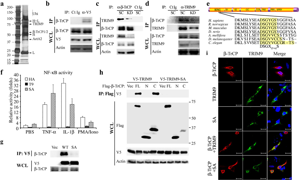
(a) Anti-V5 immunoprecipitates from 293T-V5-TRIM9 cells were separated by SDS-PAGE and visualized by silver staining. Specific bands were cut and analyzed by mass spectrometry. H, IgG heavy chain; L, IgG light chain; ActA2, actin A2. (b)Immunoblot analysis of V5-TRIM9 complexes with anti-β -TRCP. (c and d) SK-N-AS neuroblastoma cells were depleted for TRIM9 expression using scramble shRNA or TRIM9-specific shRNA lentivirus and then used for immunoprecipitation (IP) and immunoblotting (IB) with the indicated antibodies. Ct Ig: isotype control immunoglobulin. (e) Diagram of the conserved domains of TRIM9 (upper) and the degron DSGX(2+n)S motif of TRIM9 orthologs (lower). B1, B-Box1; B2, B-Box2; CC, coiled coil; COS, C-terminal subgroup one signature; FN3, fibronectin type 3; and PRY-SPRY, also known as B30.2. (f). At 24h post transfection with TK-Renilla luciferase transfection control plasmid, NF-κB reporter plasmid and TRIM9 WT (T9) or SA mutant, HEK293T cells were stimulated with PBS, PMA/Ionomycin, IL-1 β or TNF-α overnight and cell lysates were then used for dual-luciferase assay. (g) At 48h post transfection with Flag-β-TrCP and HA-TRIM9 WT or SA mutant, HEK293T cells were used for immunoprecipitation (IP) and immunoblot (IB) with the indicated antibodies. (h) At 48h post transfection with V5-TRIM9 WT or SA mutant together with Flag-β -TrCP full-length (FL), N-terminal region (N) or C-terminal region (C), HEK293T cells were used for IP and IB with the indicated antibodies. (i) Confocal immunofluorescence microscopy of Hela cells transfected with HA-β -TrCP (Green) together with HA-TRIM9 (Red) WT or SA mutant. DAPI (Blue) was used for nuclear staining. Scale bar=10µm. The data are representative of three independent experiments.