Figure 1.
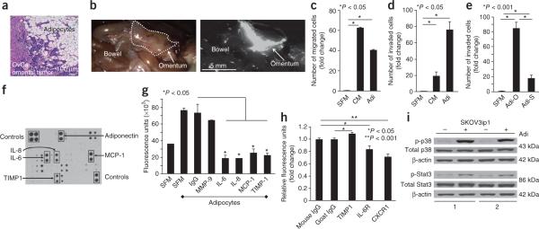
Adipocytes promote homing of ovarian cancer cells to the omentum. (a) H&E staining of ovarian cancer (OvCa) tumor cells invading adipocytes in the human omentum. (b) In vivo homing assay. Fluorescently-labeled SKOV3ip1 human ovarian cancer cells were injected intraperitoneally into nude mice. Cancer cell localization was detected after 20 min (n = 6 mice). The mouse omentum is outlined in the bright-field image (left) and visible in the fluorescent image (right). (c,d) Migration (c) and invasion (d) of SKOV3ip1 cells toward serum-free medium (SFM), adipocyte-conditioned medium (CM) and primary human omental adipocytes (Adi). Bars report mean fold change ± s.e.m. One of three experiments, each using a different human subject samples, is shown. (e) Invasion of SKOV3ip1 cells comparing primary human omental (Adi-O) and subcutaneous (Adi-S) adipocytes from the same individual. Bars report mean fold change ± s.e.m. One of two experiments shown. (f) Cytokine expression in conditioned medium from primary human omental adipocytes. One of four arrays shown. (g) Fluorescence intensity of labeled SKOV3ip1 cells that homed to Matrigel plugs containing SFM or adipocytes in the presence or absence of inhibitory antibodies. Bars report means ± s.e.m. One of three experiments shown. (h) Fluorescence intensity of labeled SKOV3ip1 cells that adhered to sections of human omentum. SKOV3ip1 cells were pretreated with a CXCR1- or IL-6R–blocking antibody, or whole omentum sections were pretreated with a TIMP-1 inhibitory antibody (n = 5 sections per group).Bars report means ± s.e.m. from one of three experiments. (i) Immunoblot of total and phosphorylated (p) p38 (Thr180/Tyr182) and Stat3 (Ser727) in SKOV3ip1 cells cultured alone (−) or with (+) adipocytes from two human subject samples for 24 h. One of three experiments shown.

