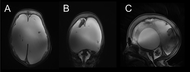
Figure 2.
(A) Transversal T2 slice across the supratentorial space with almost complete malacia of the cerebral hemispheres and with spared falx and meninges. (B) Coronal T2 slice with preserved parts of brain parenchyma in the right frontal lobe, diencephalon, mediobasal left temporal lobe, and brainstem. (C) Sagittal T2 slice with visible remnants of the diencephalon, occipital lobe, brainstem, and cerebellum. Marked thickening of the neurocranial bones is visible in all figures.

