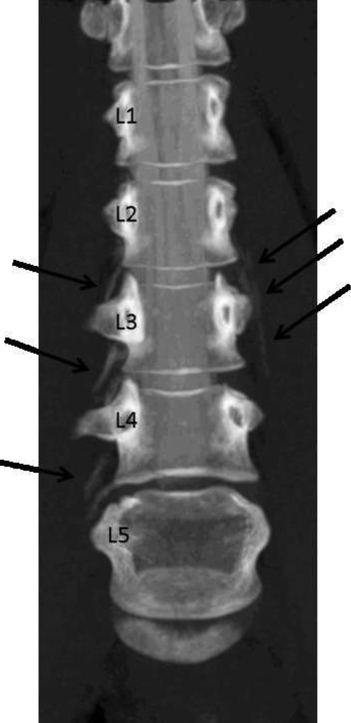
Figure 3.

After injection of the contrast agent, CT scan was performed in prone position on a Philips Brilliance 40 CT-Scanner (Hamburg, Germany) in spiral mode, collimation 40 × 0.625 mm, 120 kV, 110 mAs. Multiplanar reconstructions were calculated. Time 16:13. Maximum intensity projection in anterior-posterior view showing the lumbar spine (lumbar vertebral bodies 1-5, see numbers). The contrast agent is now visible on both sides outside the subarachnoid space distributing parallel to the spinal nerves.
