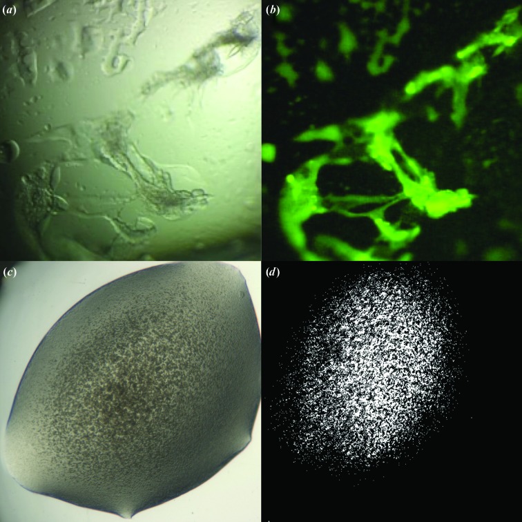
Figure 7.
Advances in the detection of crystalline material. (a) White-light image of canavalin [screening condition 30%(w/v) PEG monomethyl ether (MME) 2000, 0.2 M ammonium sulfate, 0.1 M sodium acetate pH 4.6]. (b) Fluorescence image of canavalin trace-labelled with carboxyrhodamine in the same condition. Despite the unpromising appearance of the intensely fluorescent material, optimizing around this condition led to well diffracting crystals (Pusey, 2013 ▶). (c) White-light image of a crystalline precipitate. (d) Image of the same figure taken with SONICC. Images provided by Formulatrix.

