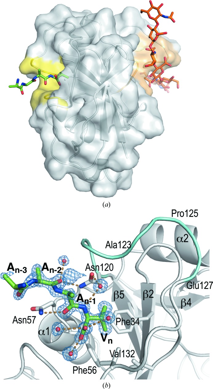
Figure 4.
The unexpected binding site for a peptide on the back side of chain B in the monoclinic hHABD18–178 structure (4pz3-B). (a) The binding site (yellow) in relation to the known HA-binding site (orange). (The HA position is modeled by superposition of the murine HABD structure 2jcr.) (b) Detail of hydrogen-bonding interactions (mostly water-mediated) and the 3σ OMIT (F o − F c) electron density (blue) validating the peptide geometry and model integrity. Backbone geometry for the three C-terminal residues (An−2, An−1 and Vn) and the side chain of the valine is very well defined. The A6 peptide homolog on CD44 (residues 120–127) is colored cyan.

