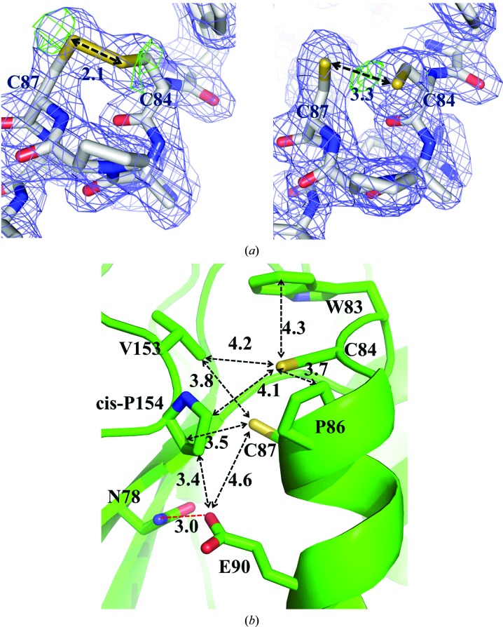
Figure 2.
Structural features of the CdDsbF active site. (a) Electron density surrounding the active-site CXXC motif of CdDsbF where the cysteines are modelled in both the (left) oxidized and (right) reduced forms. The 2F o − F c electron-density mesh (blue) and the F o − F c negative density mesh (green) are contoured at 1.0σ and 3.0σ, respectively. Shown are stick cartoons of the active site, in which the C, O, N and S atoms are coloured white, red, blue and yellow, respectively. (b) Close-up view of the CdDsbF active site showing the CAPC motif, the residues adjacent to the catalytic motif and a hydrogen-bond interaction (red dashed line) stabilizing the reduced form.

