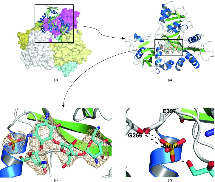
Figure 2.
The crystal structure of Msm Eis in complex with paromomycin. (a) Cartoon and surface representation of the Msm Eis hexameric structure. Five monomers of the hexamer are shown in surface mode (coloured magenta, slate, aquamarine, pale yellow and olive) and one is shown in cartoon mode. (b) Cartoon representation of the monomeric structure. (c) The electron density of paromomycin and a sulfate ion bound to chain A. The wheat-coloured mesh is the simulated-annealing OMIT F o − F c electron-density map calculated with paromomycin and the sulfate ion removed from the model (contoured at 3.0σ). (d) Cartoon representation of the sulfate-binding region in Msm Eis. Amino-acid residues and paromomycin around the sulfate ion are shown as stick models. N and O atoms are coloured blue and red, respectively. Black dotted lines denote hydrogen bonds.

