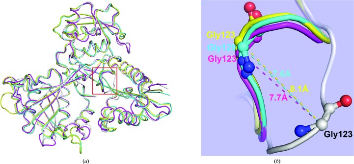
Figure 3.
Structural comparison of paromomycin-complexed Msm Eis with other Msm Eis and Mtb Eis structures. (a) Monomers of paromomycin-bound Msm Eis (grey; chain A), CoA/tobramycin-bound Mtb Eis (yellow; PDB entry 4jd6, chain A), CoA-bound Msm Eis (magenta; PDB entry 3sxn, chain A) and acetyl-CoA-bound Mtb Eis (cyan; PDB entry 3ryo, chain A) are superimposed. (b) Close-up view of the region encompassing residues Ser121–Tyr126, which are enclosed in a red box in (a), which displays a large structural difference. Amino-acid residues around this region are shown as stick models. Dotted lines denote the distance between Cα atoms.

