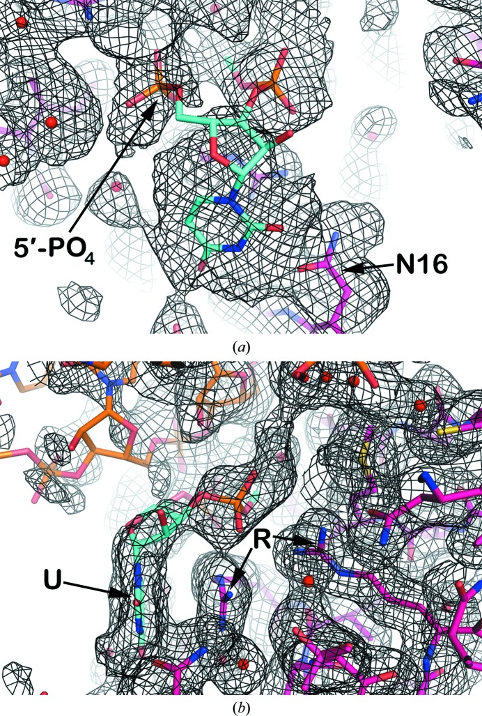
Figure 6.
The free nucleotide fragment. Displayed is the low-temperature restrained model superposed on averaged σA-weighted 2F o − F c density phased by a model which did not contain the free nucleotide. The density for both panels is contoured at 0.2σ. The free nucleotide is shown in cyan, the protein is shown in magenta, the RNA is shown in orange and water molecules are shown as red spheres. The free nucleotide is modeled with a uridine base, but the density could conceivably accommodate any base, and both the 5′-phosphate and 3′-phosphate groups are included. In (a), Asn16 is identified by ‘N16’ and can possibly interact with the uridine base through its side chain. In (b), the uridine base, identified by the symbol ‘U’, is stacked against the guanidinium group of Arg125. Arg125 and Arg131, identified by the symbol ‘R’, are hydrogen-bonded to the 3′-phosphate of the ‘free nucleotide’.

