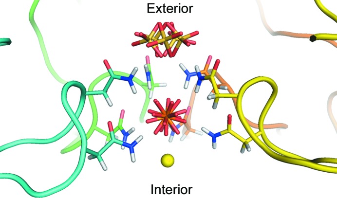
Figure 7.
The pore along the fivefold axis. The pore is viewed perpendicular to the axis. The forward-most subunit of the pentamer forming the pore has been removed for clarity. The backbone of each subunit is shown in a different color and only the side chains of the asparagine residues are shown. The Asn115 (lower residues) and Asn117 (upper residues) side chains form a cage around the phosphate ion. This ion appears to be tightly bound through ten hydrogen bonds formed by the amide N atoms of the asparagine residues. As far as we can tell, despite the disorder of the O atoms, the P atom lies on the fivefold axis. Although we originally modeled this position as a sulfate owing to the fact that the crystals were grown from ammonium sulfate, we believe that the position is probably occupied even within the plant and most probably by phosphate, which is abundant in plants. Furthermore, virus preparation involves the use of phosphate buffers that would also be a source of phosphate. Crystallographically, phosphate and sulfate are indistinguishable if both are present. At the outward side of the pore is an ion modeled as a positionally disordered sulfate because we believe that it is easily exchanged and thus is an artifact of the crystallization conditions. On the inward side of the pore we modeled a magnesium ion bound to the phosphate.

