Fig. 1.

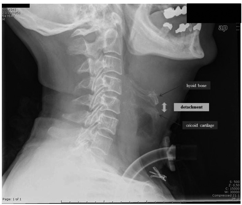
A post-operative latero-lateral neck X-ray in a patient who underwent partial reconstructive laryngectomy: the detachment of the pexy is visible as an air band between the hyoid bone (above) and the cricoid cartilage (below).

A post-operative latero-lateral neck X-ray in a patient who underwent partial reconstructive laryngectomy: the detachment of the pexy is visible as an air band between the hyoid bone (above) and the cricoid cartilage (below).