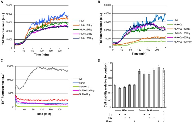
Figure 4.
Fibrillization of H6A mutated Aβ42 and scrambled Aβ42 in ThT-aggregation assay, and their cytotoxicity in rat primary cortical neurons. Fibrillization of 5 μM H6A mutated Aβ42 (H6A; light blue) (A) in the presence of increasing concentrations of Hcy (10 μM—orange, 25 μM—dark green, 50 μM—pink, and 100 μM—dark blue), (B) with 5 μM CuCl2 and increasing concentrations of Hcy (10 μM—orange, 25 μM—dark green, 50 μM—pink, and 100 μM—dark blue). (C) 5 μM scrambled Aβ42 (ScAβ; light blue) does not form fibrils when incubated alone or together with 5 μM CuCl2 or 5 μM CuCl2 + 50 μM Hcy or 50 μM Hcy. (D) Cell viability of rat primary neurons after 72 h incubation with H6A fibrils or ScAβ incubated under same conditions. Samples from aggregation assay without ThT, but with CuCl2, Hcy or both, were collected at the plateau after 4 h incubation. Concentrations were 5 μM H6A or ScAβ; 5 μM H6A or ScAβ + 5 μM CuCl2; 5 μM H6A or ScAβ + 5 μM CuCl2 + 50 μM Hcy; 5 μM H6A or ScAβ + 50 μM Hcy. As a control non-fibrillar 5 μM H6A or ScAβ (Mono) and aggregation assay reaction mixture without H6A or ScAβ were used. All values are relative to reaction mixture control sample ± S.D.

