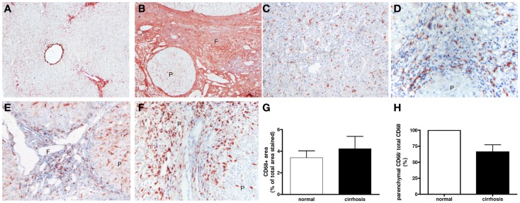
Figure 2.
Expression and localization of macrophages in normal and cirrhotic human livers. Immunohistochemical analysis shows enhanced deposition of the extracellular matrix protein collagen type I (A,B) and presence of macrophages [CD68 (C–F)] in normal (C) cirrhotic livers of various origins [(D) PBC (E) PSC and (F) congenital cirrhosis]. Note the abundant presence of macrophages in the collagenous fibrotic bands (F). (G,H) Image analysis of CD68 staining in human livers. Reduced CD68 staining was found in the parenchymal area (p) of human cirrhotic livers as compared to normal. Magnifications: 40× (A,B) and 100× (C–F). f, fibrotic matrix; p, liver parenchyma. N = 5 cirrhotic livers, N = 6 normal livers.

