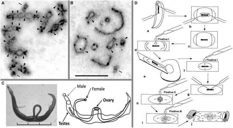
FIGURE 1.
Meiotic chromosomes, adult worms, and a technique for chromosome observation of schistosomes. Meiotic diakinesis stained by AG for Schistosoma mansoni (A) and S. japonicum (B). AG stain shows the synaptonemal complex (SC) protein, which detects crossing over between two homologous chromosomes. (C) Pairing form of the adult female and male of S. mansoni and the schematic illustration. (D) A manual preparation to obtain meiotic chromosomes from adult schistosome worms. This schema shows an example of the process with a male worm. For details, see text. Arrowhead indicates chiasma region; bar shows centriole that was stained by AG. Scale of (A,B) is 10 μm. Scale in (C) is 3 mm. See also Hirai et al. (1996, 2000) and Hirai and Hirai (2004).

