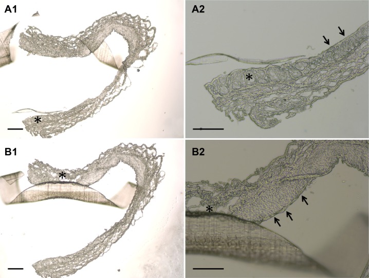
Figure 13.
Photomicrographs of cross-sections showing the condition of the stent on the vessel lumen three days following balloon angioplasty and stent deployment.
Notes: Segment of vessel of a treated animal shows a characteristic fragment of a spiral stent, wide vessel lumen without thrombus formation and non-significant stenosis without a lumen reduction of greater than 50% (A1, and B1, 40×) (scale bar: 250 mm). Additionally, segment of vessel of a treated animal has greater endothelial injury (asterisk) comparing with other vessel area (single arrow) (A2, and B2, 100×) (scale bar: 100 mm).

