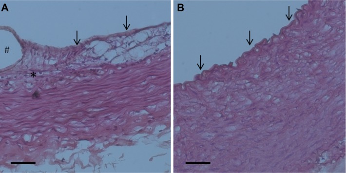
Figure 14.
Photomicrographs of representative rabbit vessel sections 28 days after intervention.
Notes: Four-week high-power photomicrographs (hematoxylin-eosin staining, ×200) of pathology of groups A and B were shown. Inflammation response was low in stent groups (A). By day 28, an anatomically intact endothelium had been re-constituted in group A (almost no intimal hyperplasia), and groups (B) exhibited consistent intimal hyperplasia with a thickness of approximately 100 μm. A single arrow represents the endothelial cells, and asterisk indicates the inflammatory cells surrounding the stent struts. Scale bar: 50 mm. # indicates the stent strut area.

