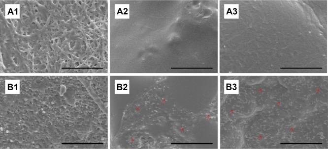
Figure 6.

Platelet adhesion test in vitro.
Notes: Following immersion of the drug-loaded and non-loaded nanofibrous membranes in platelet-rich plasma, significantly fewer platelets adhered to the drug-loaded nanofibers at 3 (A1), 7 (A2), and 14 (A3) days than to the non-loaded nanofibers (B1–3) on days 3, 7, and 14, respectively. Red arrows indicate platelet adhesion. B2 exhibits larger platelet aggregates and more extensive platelet pseudopod formation than B3 (scale bar: 50 μm).
