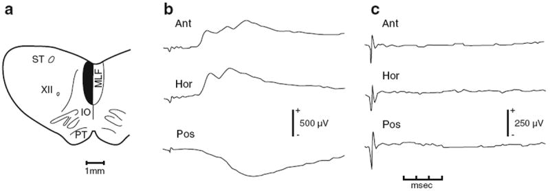
Fig. 2.

Effects of cutting the MLF ipsilateral to a dorsal neck motoneuron from which synaptic potentials were recorded in response to stimulation of contralateral ampullary nerves. ST solitary tract, XII hypoglossal nerve, IO inferior olive, PT pyramidal tract, MLF medial longitudinal fasciculus. Stimulated contralateral ampullary nerve for each trace: Ant anterior canal, Hor horizontal canal, Pos posterior canal. Lesion is shown by the dark area in a. Responses recorded in a complexus motoneuron before (b) and after the cut (c). Stimulus was 50 μA (Hor) and 75 μA (Ant and Pos) before the cut. All stimuli were 75 μA after the cut. Potentials are averages of 50 sweeps. From Wilson and Maeda (1974)
