
Figure 1. Riquiqui forms a physical complex with Dachsous and controls tissue growth.
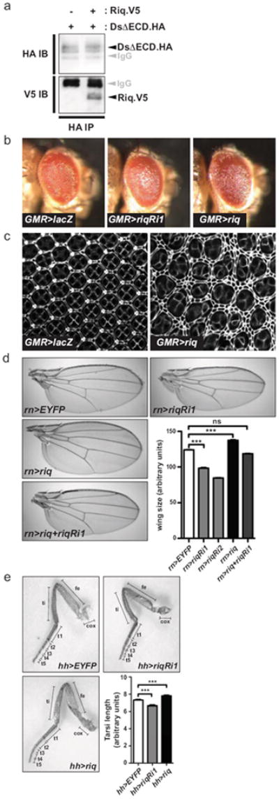
(a) Lysates from S2 cells transfected with DsΔECD.HA, alone or in combination with Riq.V5, were immunoprecipitated with anti-HA antibodies. Western blots were performed using anti-HA and anti-V5 antibodies to reveal Ds and Riq, respectively. (b) Lateral views of adult female eyes (anterior is to the right) expressing UAS-lacZ, UAS-riqRi1 or UAS-riq under the control of GMR-Gal4. (c) Eyes 44 hours APF expressing UAS-lacZ or UAS-riq under the control of GMR-Gal4 that have been stained with anti-Discs-large antibody. (d) Representative images of wings from adult female flies expressing UAS-EYFP, UAS-riqRi1, UAS-riqRi2, UAS-riq or UAS-riqRi1 concomitantly with UAS-riq under the control of rn-Gal4. Wing size was quantified for each genotype (n=20 for each). (e) Right rear legs from adult female flies expressing UAS-EYFP, UAS-riqRi1 or UAS-riq under the control of hh-Gal4. Legend of leg segments is as follows, coxa (cox), femur (fe), tibia (ti), tarsal segments 1 to 5 (t1, t2, t3, t4, t5). The length of the entire tarsal segment was quantified (n=15 for each genotype). In d and e, data are presented as mean ± SEM, *** = p<0.001, ns = non-significant.