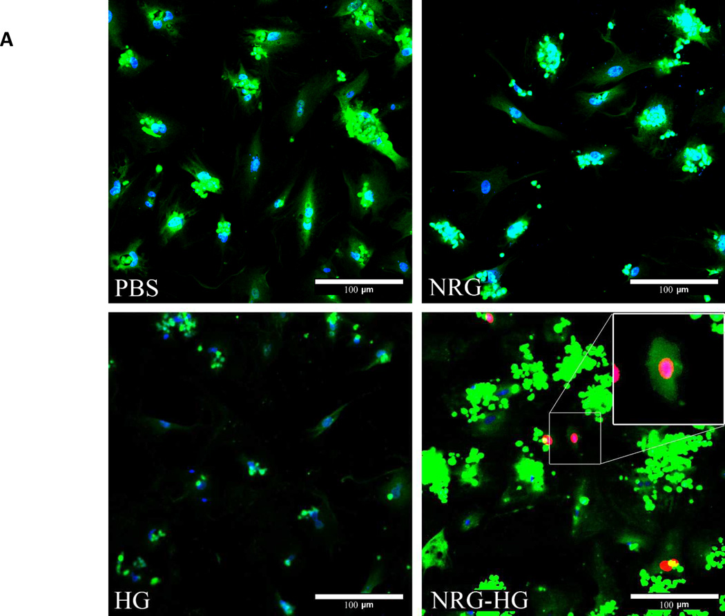
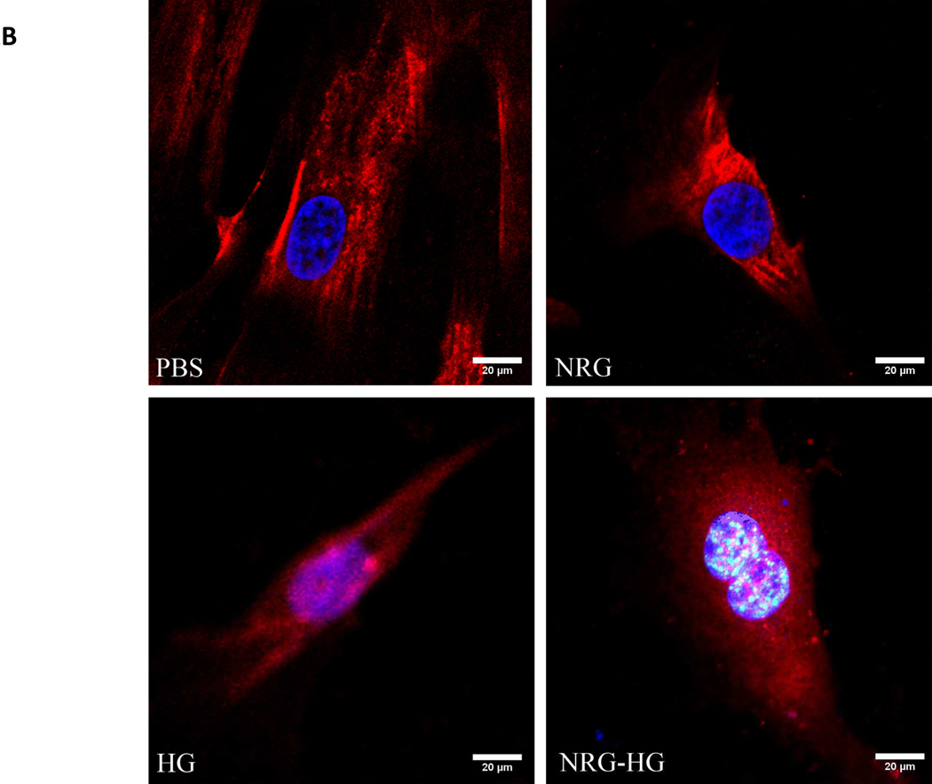
Figure 2. Cardiomyocyte mitotic activity.
Confocal microscopy illustrating EdU incorporation and PH3 in isolated cardiomyocytes 6 days following stimulation. Cells are stained with DAPI (blue), troponin (green), and EdU (red). 10% of NRG-HG treated cardiomyocytes were positive for EdU incorporation. A higher magnification view is provided for the EdU positive cardiomyocyte (A). For PH3 staining, cells are stained with DAPI (blue), troponin (red), and PH3 (green). 2% of NRG-HG treated cardiomyocytes were positive for PH3 (B).


